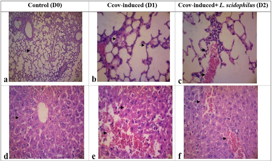
Figure 2:
The histological examination of the control group (a, d) demonstrates typical cell structure in the lung tissues and liver. In the D1 group (b), lung tissues exhibit histomorphological feature such as diffuse alveolar cell damages, hyaline tissues deposition, septa alveolar hemorrhages, leukocyte cells infiltration, and alveolar septa proliferation. The D2 group (c) shows neutrophil cell infiltration and plasma fibrin accumulation in lung tissues. In the liver tissues of D1 group (d) shows an accumulation of glycogen in hepatocyte cells, hemorrhage with dilated sinusoids, infiltration of lymphocyte cells. The D2 group (e) present sinusoidal congestion and dilatation (HE, 400x, changes are indicated by arrows).