
Fig. 1.
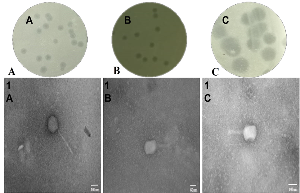
Plaque morphologies of phage SEPL01 with a halo (A), SEPL13 (B) and SEPL20 (C). Electron Micrographs of phage SEPL01 (1A), SEPL13 (1B), and SEPL20 (1C).
Plaque morphologies of phage SEPL01 with a halo (A), SEPL13 (B) and SEPL20 (C). Electron Micrographs of phage SEPL01 (1A), SEPL13 (1B), and SEPL20 (1C).