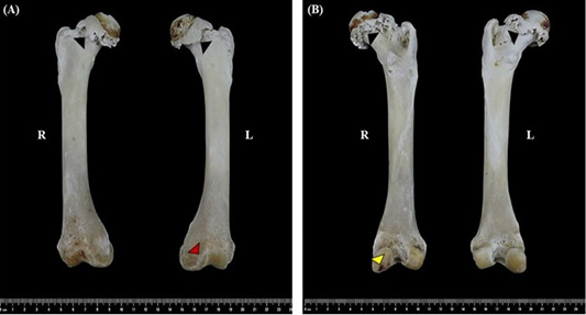
Figure 1
Osteophyte formation along the femoral head-neck junction (black arrowheads) and the trochlear margin of the distal femur (red arrowheads). The right femoral head showed a flattened shape, compared with the rounded appearance observed in the left femoral head. Evidence of subchondral bone degradation was observed on the surface of the right medial femoral condyle (yellow arrowheads). (A) cranial view of right (R) and left (L) femur. (B) caudal view of right (R) and left (L) femur.