
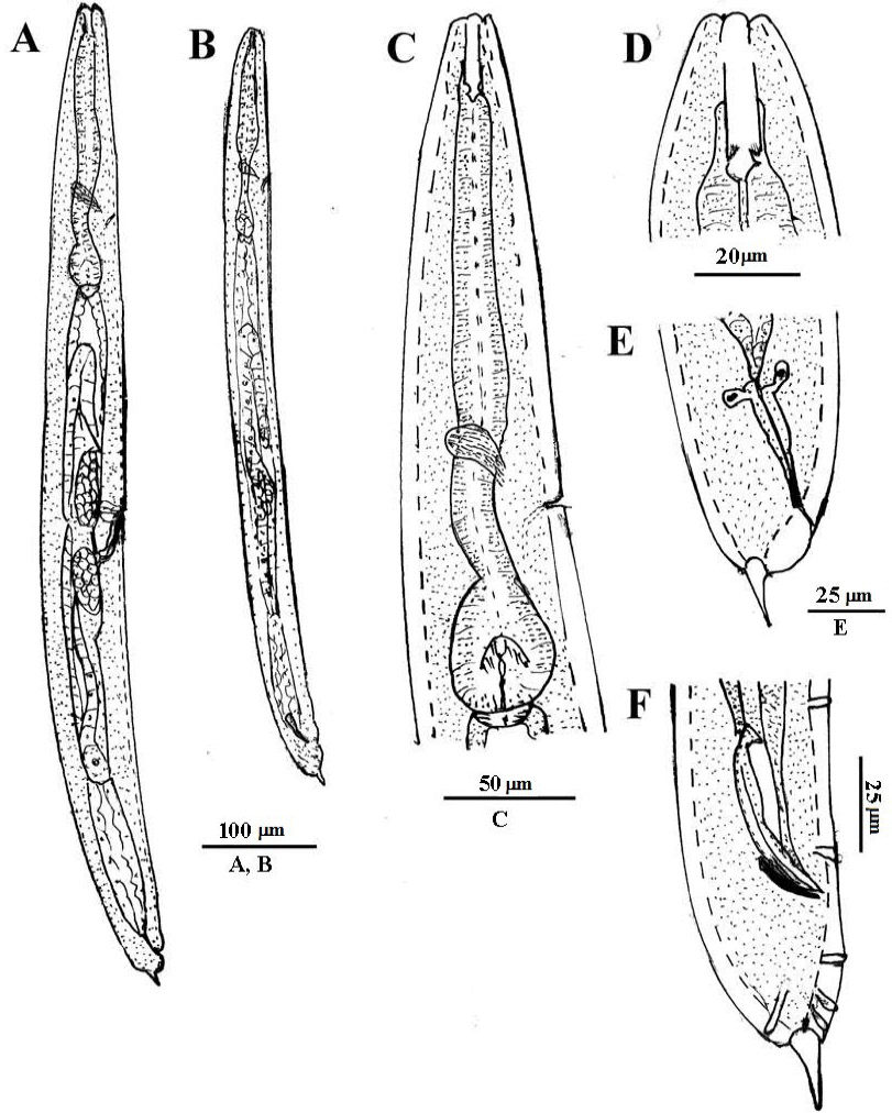
Fig. 2.
Poikilolaimus oxycercus de Man, 1895 (A-F). Female (A,C-D): A, whole body; C, pharyngeal region; D, anterior region; E, tail region. Male: B, whole body; F, ventral view of tail region showing papillae.
Poikilolaimus oxycercus de Man, 1895 (A-F). Female (A,C-D): A, whole body; C, pharyngeal region; D, anterior region; E, tail region. Male: B, whole body; F, ventral view of tail region showing papillae.