
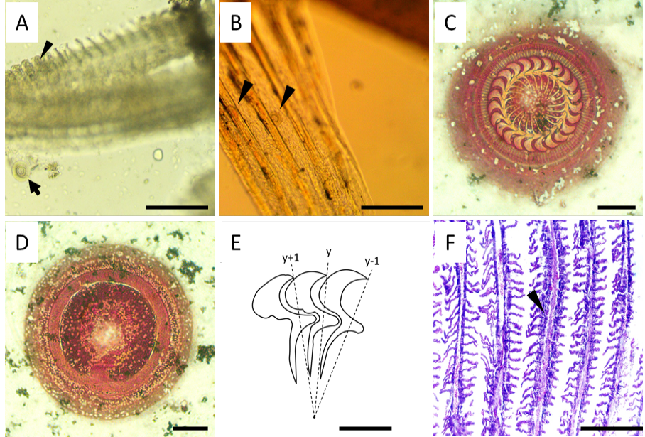
Figure 1:
Trichodina heterodentata parasitizing Piaractus brachypomus. (A) Fresh-mounted smear from the gill arches showing light hyperplasia (arrowhead) and Trichodina (arrow); (B) Trichodinids (arrowheads) in a fresh-mounted smear from the fins; (C) Silver impregnated adhesive disc; (D) Nuclear apparatus; (E) Schematic drawing of the denticles; (F) Histological section of gill from parasitized fish showing light hyperplasia (arrowhead) and thickening of gill filaments. H-E. Scale bars: A, B, F = 200 µm; C, D = 10 µm; E = 5 µm.