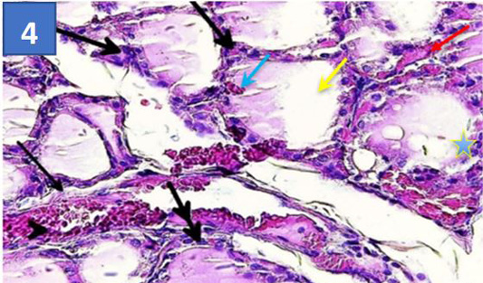
Figure 4:
Section of thyroid gland treated by SAL: Showing thickness of tissue seprated of the lobules (black darts)moderete depletation of para follicular cell (thick arrows), scanty colloid (yellow dart) hypermia between thyroid follicles(red dart),and between thyrocyte (blue dart), infiltrated of inflmmatory cell around some follicles (double arrow), hyperplastic capillaries (arrow head) and intrafolliculor hyperplasia (blue star) (40X).