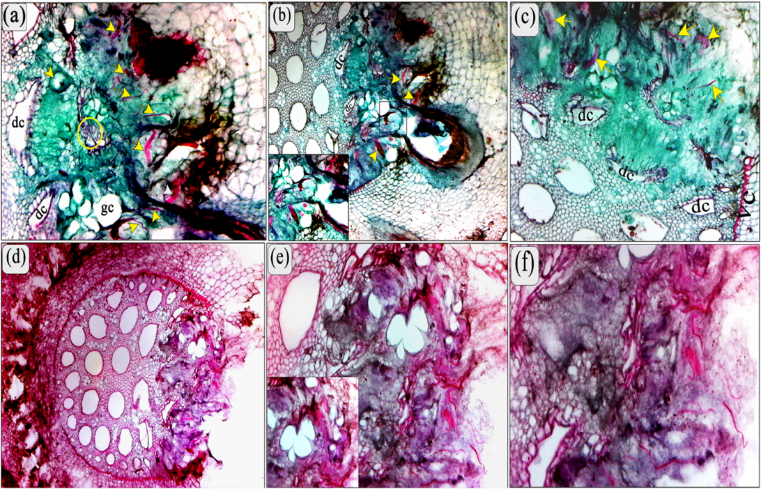
(a) Transverse section of banana roots showing the adult female of M. incognita (arrows) inducing the clear multinucleate cells (cycle) and with fused syncytial cells presenting dense cytoplasm and hypertrophic nuclei, and shows some of adult and larval stages of R. similis (arrows). (b) Showing deformed cells (dc) and noted that the extension of the feeding site to the vascular cylinder. High power showing the most damage in root cells as results of a combination of both nematodes. (c and d) root infested with pre-adult stages of both M. incognita and R. similis showing the abnormal xylem, clear dissolution of endodermis and central cylinder (arrows) and note the thickness of endodermis layer compared to the normal size in the control. Also, note all the modification symptoms associated with the feeding process; multinucleate cells, giant cells, and modified cells. (e, f and d) The laceration and dissipation of the central cylinder (xylem and phloem vessel) as a result of the feeding of both species. Note the extent of the feeding sites inside the vascular cylinder. (Scale bars: a-f = 200 μm).