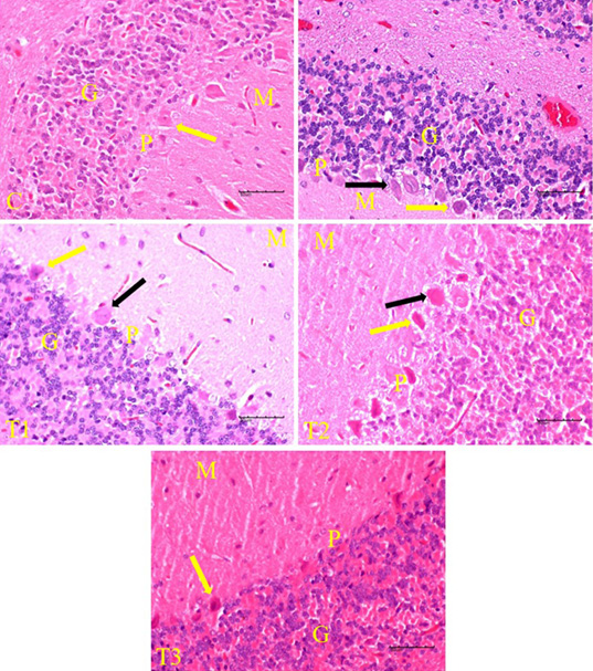
Figure 2:
Histological changes in the cerebellar cortex of rats exposed to LA and treated with EBN. Note that darkly stained degenerating pyramidal cells (black arrow) among the healthy pyramidal neurons (yellow arrow) in T0, T1 and T2. Cerebellar layers include molecular(M),purkinje(P) and granular (G).