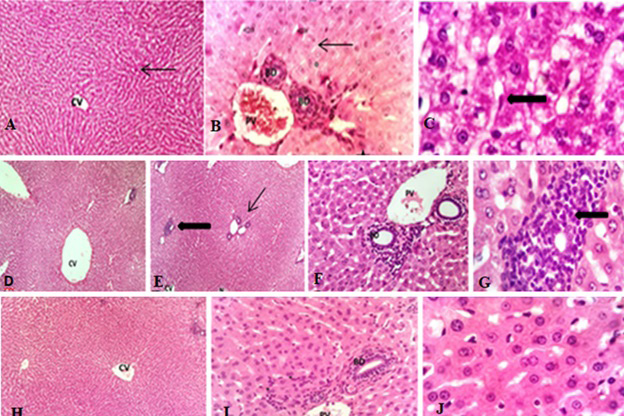
Fig. 1.
Effect of propranolol on histological structure of phenytoin-treated liver of albino rabbits.
A, Photomicrograph of 5 micron thick hematoxylin and eosin (H and E) stained paraffin section from liver of rabbits of control group showing normal central vein (CV) and radiating hepatic architecture and normal sinusoidal spaces (arrow)(100X). B, showing normal portal vein (PV) and bile duct (BD), normal sinusoidal spaces (arrow) and binucleated hepaatocytes (star) (400X). C; showing normal hexagonal hepatocytes, kupffers cells (thick arrow) (1000X). D, PHY treated group showing moderately disturbed hepatic cords with dilated sinusoids especially in periportal area. The central vein (CV) was also markedly dilated (100X). E, showing necrotic patches (N), polymorphonuclear cell infiltration (thick arrow) especially within portal tract and hemorrhagic sinusoids (thin arrow) 100X. F, illustrating that the portal tract was severely inflamed with mononuclear cells with dilated and congested portal vein (PV) (400X). G, pointing out the inflammatory patch. Plasma cells and neurophils were observed within the patch (thick arrow) (1000X). H, PHY and PRL treated group showing almost normal hepatic lobules with normal uninflamed central vein (CV) (100X). I, showing small number of inflammatory cells in portal tract. The diameter of portal vein (PV) mildly increased (400X). J, explaining that the hepatic cells were normal with granular cytoplasm and nucleus with nucleolus 1000X.
