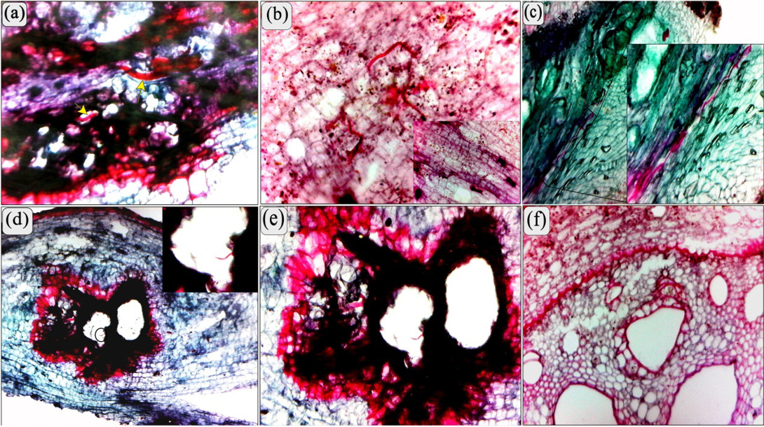
Figure 1:
(a, b, and c) Longitudinal section of a banana root showing pre-adult stages and adult stage of Radopholus similis (arrow) coiled in medial parenchyma. (b) High power represents the lignification in sub-epidermal cortical cells. (c) High power showing the pathway of pre-adult stages of R. similis in the cortical parenchyma. (d and e) showing the overlap infection between pre-adult stages of Radopholus similis through the gall formed by M. incognita. (f) Transverse section of a banana root showing the modified xylem, phloem vessels and parenchyma caused by Radopholus similis. Note a few of phenolic and lignified cells in vascular cylinder (b). (Scale bars: a = 70 μm; b-f = 200 μm).