
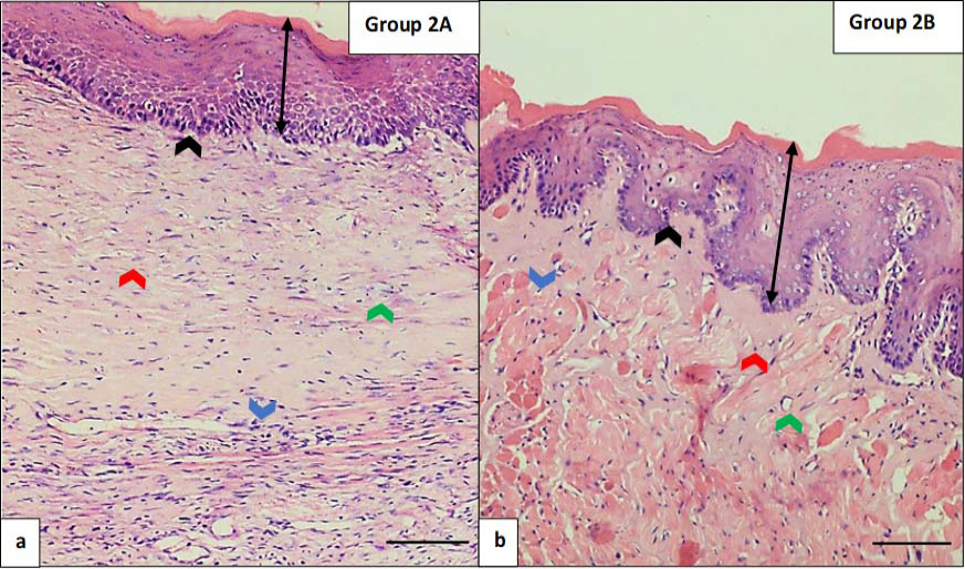
Fig. 3.
Effect of muco-adhesive gel on oral submucous fibrosis in buccal mucosa of rat after 12 weeks of treatment.
(a) Experimental group 2A at week 12 (scale bar=100 µm). (b) Experimental group 2B at week 12 (scale bar=100 µm). In the 12th week, the experimental group 2A (a) showed advanced stage OSMF features i.e., thin and atrophic epithelium with orthokeratosis (double black arrows), shallowed and absent rete pegs (black arrows), vascular constriction (green arrow), reduced cellularity, dense collagen in the form of sheets in connective tissue (red arrows) and muscular atrophy (blue arrows). In the 12th week, the experimental group 2B (b) showed moderate stage OSMF features i.e., varying thickness of epithelium with orthokeratosis (double black arrows), broad rete pegs (black arrows), dilated vasculature and vascular occlusion disappearance, few inflammatory infiltrate, dense collagen in connective tissue just below the epithelium (red arrows) and muscle fibers interspersed among collagen deposits (blue arrows) (Stain: hematoxylin and eosin, Magnification: 10X).