Figure 3
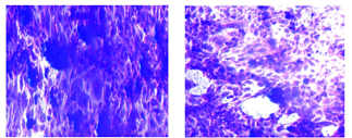
Microscopic images show morphological alteration of CAL-51 cell lines. (left) treated with AGE, (right) untreated cells. The cells were stained with crystal violet (20X).
Microscopic images show morphological alteration of CAL-51 cell lines. (left) treated with AGE, (right) untreated cells. The cells were stained with crystal violet (20X).