
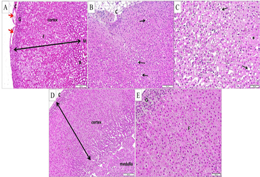
Figure 4:
(A, B and C) Adrenal gland from depression group (H and E stain A and B X200 and C X400, respectively), showing. (A) Cortical zones; glomerulosa (G), fasciculata (F) and reticularis (R) and the medulla (M), with apparent increase of cortical thickness and apparent intracapsular cavities (red arrows). B) Disorganization of cortical zones and fat deposition in the capsule (c). (C) Hypertrophied cells in zona fasciculata with large cytoplasmic vacuoles (black arrows). Some cells show absent nuclei (*). (D and E) Adrenal gland from the depression exercise group (H and E stain X200and X400 respectively) with restoration of the normal adrenal architecture and normal cellular organization in zona glomerulosa (G) and fasciculata (F).