Figure 7:
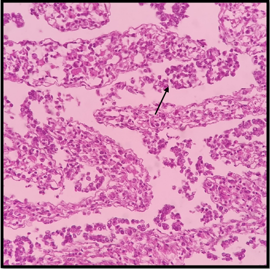
G2 (23 DPI) the mucosa and submucosal tissues were severely disrupted, and there were marked cystic crypt dilations and disorganized villus structures. A higher magnification of the previous section revealed the complete loss of enterocytes along with remnants of proprial tissue that still contained severe inflammatory cells, necrotic debris between villi (black arrow), (H & E stain X 40).