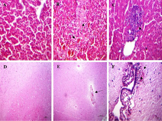
Histopathological lesions in the liver and brain of naturally infected quails; (A) liver of normal quail showing normal hepatic parenchyma, (HE, x400); (B) liver of infected quails with NDV showing intense sinusoidal congestion, large area of coagulative necrosis containing erythrocytes apoptosis(arrow) and minute necrosis of the hepatic parenchyma, (HE, x400); (C) liver of infected quails with NDV showing interstitial lymphocytic aggregation, (HE, x400); (D) brain of normal quail showing apparently normal brain tissue, (HE, x100) (E) brain of infected quails with NDV showing perivascular hemorrhages in Virchow robin spaces and non-suppurative encephalitis. besides, hemorrhages in meninges, (HE, x100); (F) brain of infected quails with NDV showing sub meningeal lymphocytic infiltration, extra vasated erythrocytes and edema, (HE, x400).