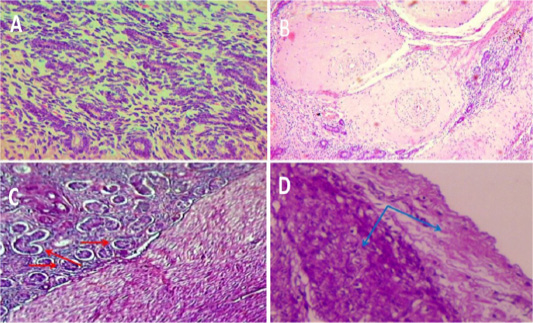
Figure 4:
A: Chronic endometritis: uterine gland showing Atrophy of endometrial glands with infiltration of inflammatory cell in lamina propria (400x H & E). B: Chronic endometritis: diffuse of Medial hypertrophy of blood vessels with stenosis of lumina and infiltration of inflammatory cells in lamina propria (100x H & E). C: Adenomyosis: uterine glands along with adjacent stromal tissue within myometrium (red arrow). 100x H & E. D: Metritis: the presence of inflammatory cells and edematous fluid within the myometrium and serosa (blue arrows). 100x H & E.
