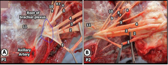
Fig. 3.
Gross Anatomical image of the brachial plexus and its branches. A shows determines the anatomical dissection of the root of the brachial plexus at site P1 with the appearance of an axillary artery, while B shows determines the anatomical dissection of the brachial plexus after bifurcation from the origin at site P2.
Abbreviations: 1, musculocutaneous nerve; 2, median nerve; 3, the ulnar nerve; 4, radial nerve; 5, axillary nerve; 6, lateral thoracic nerve; 7, suprascapular nerve; 8, subscapular nerve; 9, caudal pectoral nerve; 10, dorsal thoracic nerve; and long 11, thoracic nerve.
