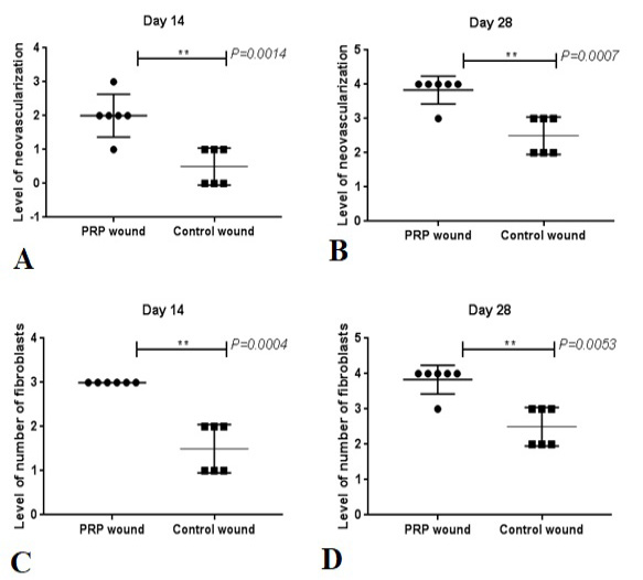
Fig. 4.
Histopathological scoring of neo-vascularization and fibroblast between the both groups. A, indicates level of neovascularization in PRP and Control wounds at day 14. B, indicates neovascularization in both groups at 28 days. C, indicates level of number of fibroblasts in PRP and Control wounds at day 14. D, indicates level of number of fibroblasts in both groups at 28 days. **, indicated the differences were significant in the PRP-treated group compared with the control group at day 28 (P<0.01).