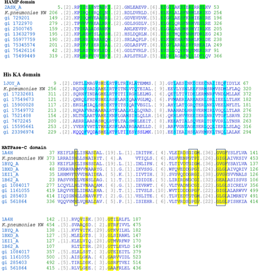
Fig. 6.
Multiple alignment of the three domains in CusS of K. pneumoniae KW along with related domain sequences found in CDD. In HAMP domain, residues involved in dimerization interface are highlighted with green colour. In His KA domain, phosphoacceptor site and residues involved in dimerization are highlighted with yellow and blue colours, respectively. In HATPase-C domain, residues involved in ATP binding are highlighted with yellow colour while Mg binding site and GXG motives are boxed with blue and black boundaries, respectively.
