
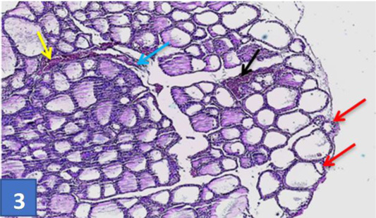
Figure 3:
Section of thyroid gland treated by SAL: showing atrophied with shrinking follicles (red darts), congested large blood vessel (yellow dart), and hyperplastic changes (black dart) thickness of connective tissue separated the thyroid lobules (blue dart) (H & E) stain (10X).