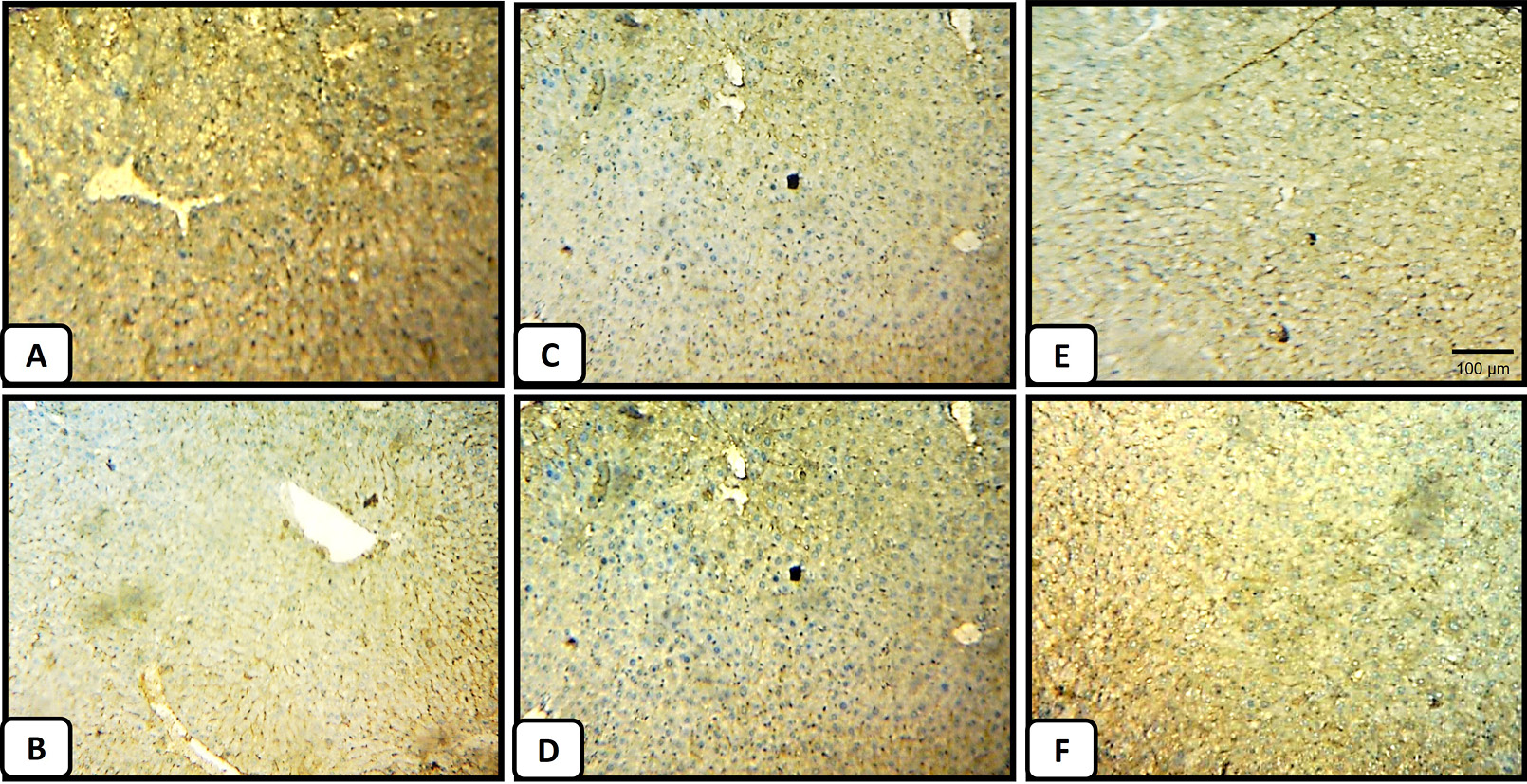
Immunohistochemistry of the cytoprotective molecule, Bcl-2, in the liver tissues. (A) A representative section from a negative control mice liver shows a normal pattern of Bcl-2 with mild staining. (B) The liver section obtained from CCl4-intoxicated mice shows faint staining (indicated by brown color) for Bcl-2. (C) Liver section from normal mice treated with M. forsskalii extract at a low dose shows a detectable reduction in the expression of Bcl-2 in the normal liver when introduced at a low dose (Magnification, x 50), (D) A representative liver section from normal mice treated at high dose shows mild staining ability by Bcl-2, indicating a very limited effect of M. forsskalii extract on Bcl-2 expression when administered at high dose, (E) and (F) Liver tissue sections prepared from the 100 mg/kg and 500 mg/kg of M. forsskalii after CCl4 treatment groups, respectively, showing the relative restoration of the Bcl-2 staining compared to (B), however (F) shows closer staining pattern to the normal liver (A).