
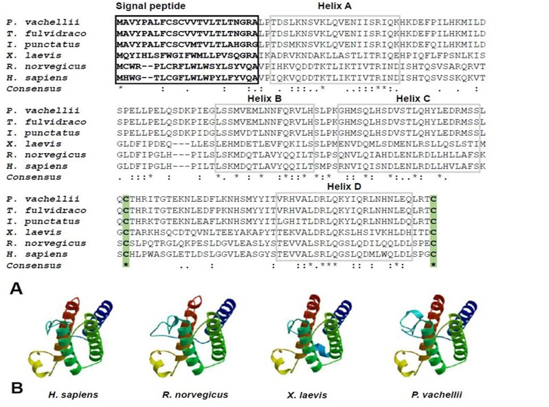
Fig. 2.
Multiple Leptin sequences alignment (A) and three-dimensional structure modeling of Leptins in four representative species (B). Signal peptide sequences are marked by black box and highlighted with bold font. Four α-helix domains were labeled by gray boxes and numbered A-D. Two conservative cysteine residues were marked by green background with bold font. The representative species including human (H. sapiens), rat (R. norvegicus), frog (X. laevis) and darkbarbel catfish (P. vachellii).