
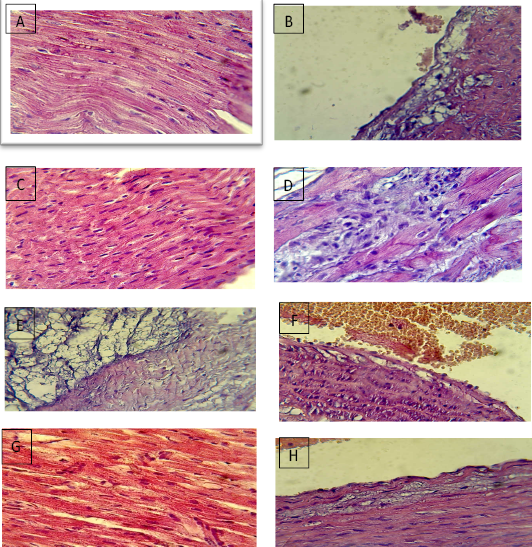
Figure 1:
Section in the heart of I+D group shows no clear lesions (A). Section in aorta of I+D group shows vacuoles in subintima (B). Section in the heart of I+A group shows no clear lesions (C). Section in the heart of I+A group shows no mononuclear cells infiltration between cardiac muscle (D). Section in the heart of 0+D group shows necrotic cardiac (E). Section in the aorta of 0+D group shows vacuolation in subintima (F). Section in the heart of 0+A group shows no clear lesions (G). Section in the aorta of 0+A group shows no clear lesions (H).