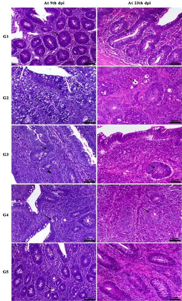
Histological structure of intestine (cecum) showing normal both covering epithelium and intestinal crypts (arrow) at 9th and 23th dpi., in non-infected non-treated chicken (G1), heavily infestation of the intestinal crypts with different coccidial stages (arrow) at 9th dpi. and dead coccidial stages (arrow) at 23th dpi., in infected non treated chicken (G2), marked decrease of affected crypts which contained the schizont of the parasite (arrow) and associated with intestinal hyperplasia (arrowhead) at 9th dpi. and hyperplasia of the crypts lining epithelium (arrow) with mild interstitial mononuclear cells infiltration (arrow) at 23th dpi., in infected chicken treated with cashew oil (G3), marked decrease of the number of live coccidial schizont stages (arrow) at 9th dpi. and hyperplasia of the crypts lining epithelium (arrow) with slight interstitial mononuclear cells infiltration at 23th dpi., in infected chicken treated with toltrazuril (G4), normal mucosa with apparent dead parasitic stages within the intestinal epithelial cells (arrow) at 9th dpi. and slight hyperplasia of the crypts lining epithelium (arrow) at 23th dpi., in infected chicken treated with cashew oil + toltrazuril (G5), H & E, X200.
