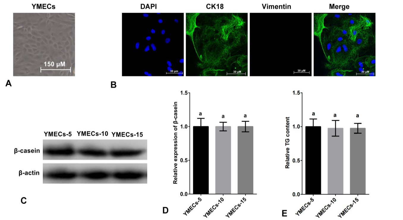
Fig. 1.
Identification of purified YMECs. A: Cell morphology (100×). B: Expression of CK18 in YMECs (400×). Cell nucleus were dyed with DAPI (blue), CK18 was visualized with Alexa Fluor 488 conjugated antibody (green) and vimentin (a marker of fibroblast, used as negative control) was visualized with Alexa Fluor 647 conjugated antibody red. C-D: Expression of β-casein in YMECs within 15 generations. E: Secretion of TG of YMECs within 15 generations. YMECs-5, 10 or 15: YMECs were 5, 10 or 15 generations, respectively.