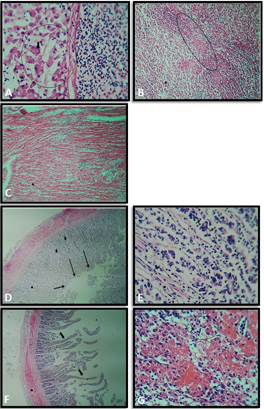
Fig. 4.
Histo-pathological changes in liver (A, B), broiler heart (C), intestines (D, E,F) and spleen (G).
The encircled area in A (positive control) shows necrosis with empty spaces along with karyorrhexis and diffused congestion in the liver. The encircle area in B shows the diffused congestion in the liver. C Shows extensive presence of inflammatory cells in the myocarditis. D shows disrupted villi (arrows) while the stars in E indicated elongation of the glands of the cecum and lymphocytic proliferation (arrows) in cecum of W. coagulans group Photomicrographs of broiler intestine and spleen group 4. F showes abnormal elongation of the villi of intestine of Nigella sativa treated group encircled area in G showed the infiltration of inflammatory cells, lymphocytes and neutrophils in spleen.
