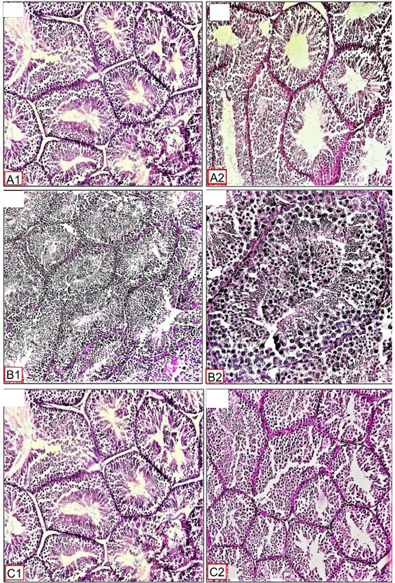
Fig. 6.
Histological photomicrograph structure of testis of testis of TT50, SPT+TT100 and SPT+TT50 treated pigeons under a long photoperiod (19L:5D) showing: TT50 croup (A1 and A2) Normal testicular tissue, normal seminiferous tubules, and a higher number of Spermatozoa and epithelial germ cells are observed. Our examination revealed that SPT+TT100 (B1 and B2) and SPT+TT50 (C1 and C2) groups showed a testis tissue that seemed close to the control. The extract has achieved in partially decreasing the damage induced by SPT. Magnification: A1, B1, C1, C2 = X10. A2, B2 = X40. Stain H and E.