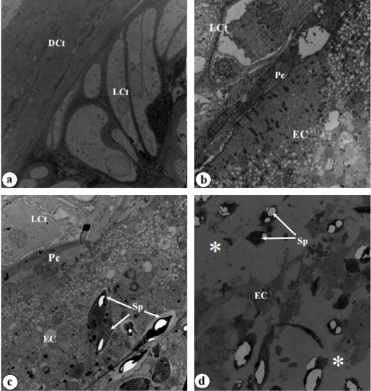
Figure 6:
Transmission electron microscope photographs of the tip of the ABO bulb. (a) Section displaying the dense connective tissue (DCt) covering the loose connective tissue (LCt) (x 2,500). (b) Section revealing the loose connective tissue (LCt) and the elastic cartilage (EC), with the perichondrium (Pc) separating the loose connective tissue (LCt) from the elastic cartilage (EC) (x 2,000). (c) Parasite-host interface demonstrating myxospores (Sp) within the elastic cartilage (EC), with the perichondrium (Pc) undergoing significant damage (x 2,200). (d) Section of the elastic cartilage (EC) containing myxospores (Sp), displaying cellular destruction within the elastic cartilage (EC) and empty spaces (white stars) (x 2,500).
