
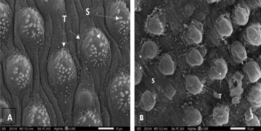
Figure 3:
Scanning electron micrographs of S. mansoni worm collected from (A) Infected control mouse showing normal dorsal tegument tubercles (T) with spines (S). (B) Infected mouse treated with FWE showing mostly reduced spines (S) density and abnormal tubercular shape (T).