
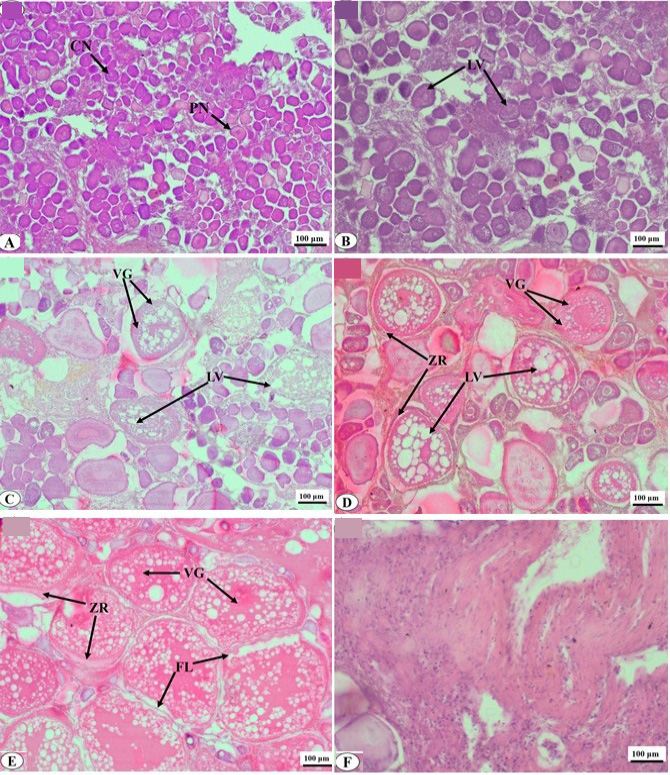
Fig. 1.
Histological preparations of oocytes of silver pomfret (A) primary growth; chromatin (Cn) and the perinucleolus stage (Pn) (B) yolk vesicle, LV, lipid vesicles (C) early vitellogenis. VG, Vitellin globules (D) Late vitellogenis. ZR, zona radiata (E) mature stage. FL, follicular layer (F) postovulatory follicle stage (Pof).