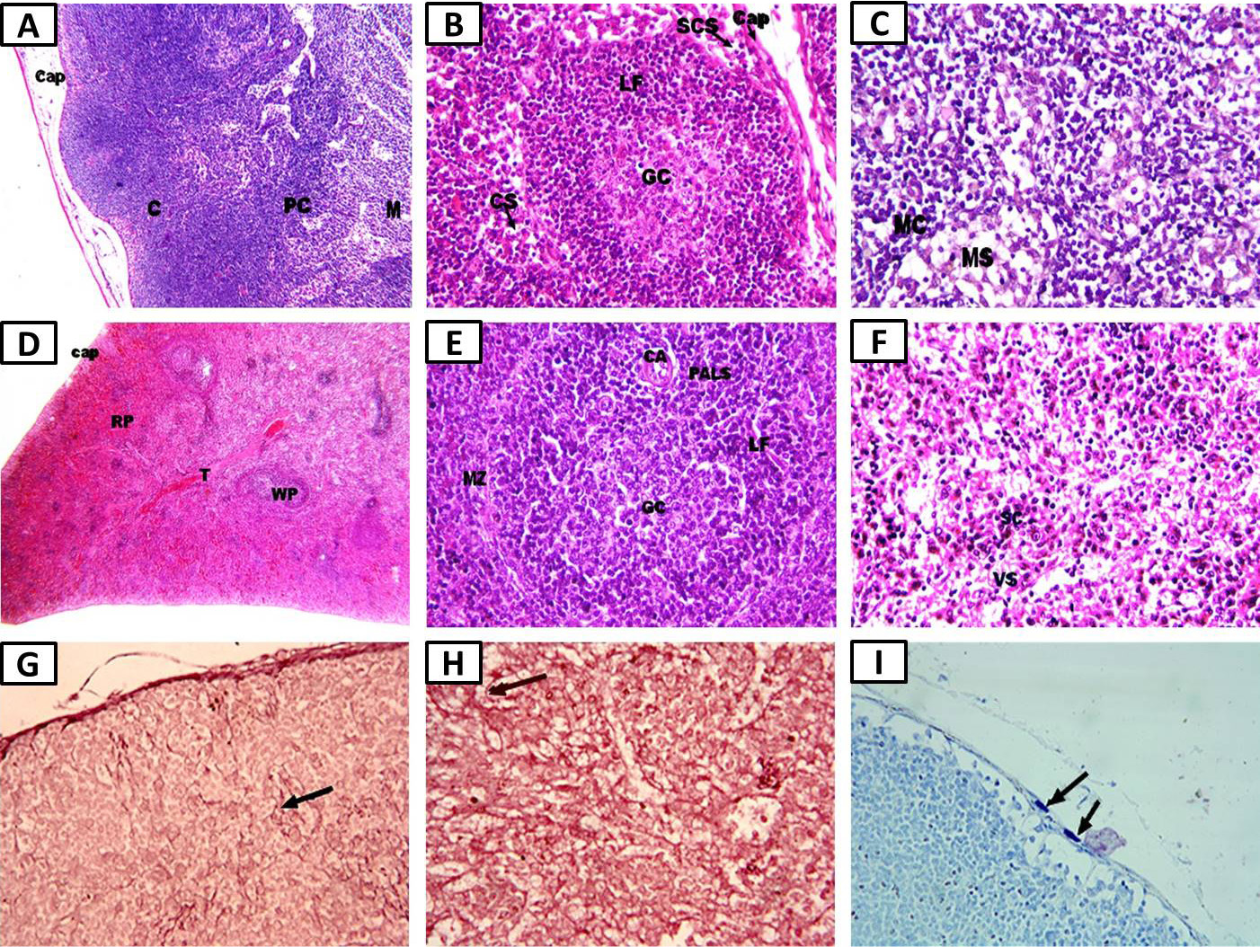
Photomicrographs of the negative control group showing: A, normal mesenteric lymph node structure with cortex (C), paracortex (PC), and medulla (M) (×100); B, cortex with lymphoid follicle (LF), germinal center (GC), capsule (Cap), cortical sinuses (CS), and subcapsular sinus (SCS) (×400); C, medulla with medullary cords (MC) and medullary sinuses (MS) (×400); D, normal spleen structure with white pulp (WP) or lymphoid follicles (LF), vascular red pulp (RP), outer capsule (C), and trabeculae (T) (×100); E, white pulp’s LFs with germinal center (GC), periarteriolar lymphatic sheath (PALS), central arteriole (CA), and marginal zone (MZ) (×400); F, red pulp (RP) with splenic cords (SC) and venous sinuses (VS) (hematoxylin and eosin, ×400); G, normal thin delicate elastic fibers in lymph node cortex (arrow) (×400); H, normal thin delicate elastic fibers in spleen RP (arrow) (Orcein, ×400); I, normal number of flattened mast cells (arrows) in the pericapsular connective tissue of the lymph node (toluidine blue, ×400).