
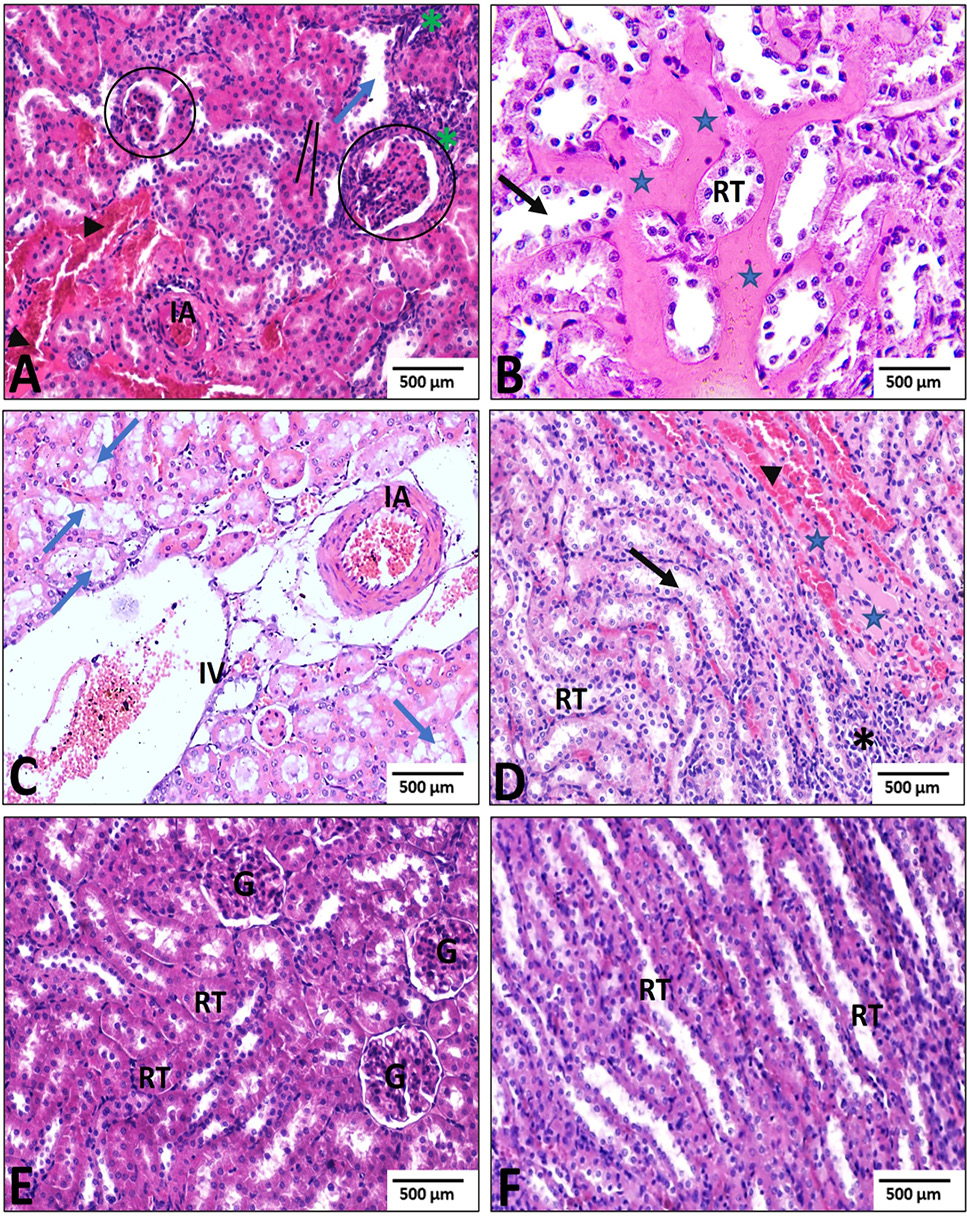
Fig. 2.
Effect of aqueous myrrh extract (AME) on histological structure of Ethanol (EtOH) and EtOH + AME-treated groups. A–D, Renal tissue of rats from the EtOH-treated group showing shrunken glomeruli (circle), inflammatory infiltration (*), tubular cell necrosis with vacuolization (blue arrow), dilation of tubular lumens (black arrow), interstitial edema (star), tubular congestion (arrowhead), pyknotic nuclei in most epithelium lining the tubules (line), and expansion of interlobular veins (IV). E and F, Renal sections of rats treated with EtOH + AME showing improved histopathological changes in the renal cortex (E) and medulla (F). G, glomeruli; RT, renal tubule. Scale bar =500 µm; Stain: H & E. Magnification: 200 and 400 ×.