View larger version:
Download Original File
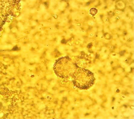
Figure 1:
Micrograph of the hemosome form of light microscope 100 x.