
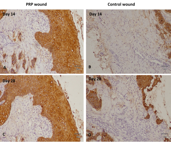
Fig. 5.
Cytokeratin staining in tissues obtained at Days 14 and 28 after wounding for Re-epithelization in both groups. A, PRP gel treated wound tissue displays accelerated epithelial growth, increased proliferation of connective tissue at day 14. B, Control wound tissue displays no development of epithelial growth, no basal lamina growing, and mild proliferation of connective tissue at day 14. C, PRP gel-treated wound tissue displays increased epidermal growth, intact basal layer, multiple layers of stratum spinosum visible, stratum corneum also visible, and increased fibrous connective tissue on day 28. D, Control wound tissue mild epithelial growth, basal lamina growing, and mild proliferation of connective tissue at 28 days.