
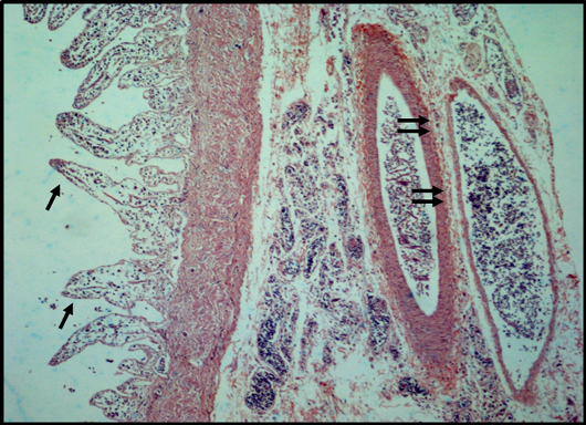
Figure 3:
Photomicrograph of intestine showing an increase in apparent size of villi with blunt and pointed ends (arrow), shrinkage of serosa and muscularis mucosa along with necrotic patches can be seen visible and nerve plexus (double arrow).