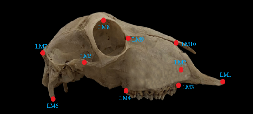
Fig. 2.
Lateral landmarks: LM1, Anterior edge of incisiv bone; LM2, Infraorbital foramen; LM3, Anterio-dorsal edge of PM1; LM4, Caudal edge of M3; LM5, Middle point of zygomatic arch; LM6, External acoustic pore; LM7, Ventral edge of jugular process; LM8, External occipital protuberance; LM9, Ventral edge of occipital condyle; LM10, Middle point of margo supraorbitalis; LM11, Medial angle of orbita; LM12, Fissura nasomaxillaris; LM13, Anterior edge of septal process.
