
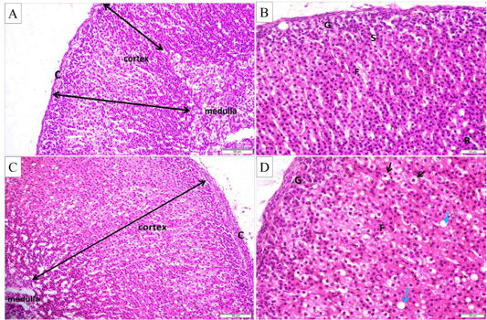
Figure 3:
(A and B) Histological structure of adrenal gland in control group (H and E stain X200 and X400 respectively), showing the capsule (c), cortex and medulla. The cortex is formed of three zones; zona glomerulosa (G), fasciculata (F) and reticularis (R). Notice the blood sinusoids (s). (C and D) Histological structure of adrenal gland in the control exercise group (H and E stain X200 and X400, respectively), showing apparent increase of cortical thickness with hypertrophied cells (black arrows) and large cytoplasmic vacuoles (green arrows) in zona fasciculata.