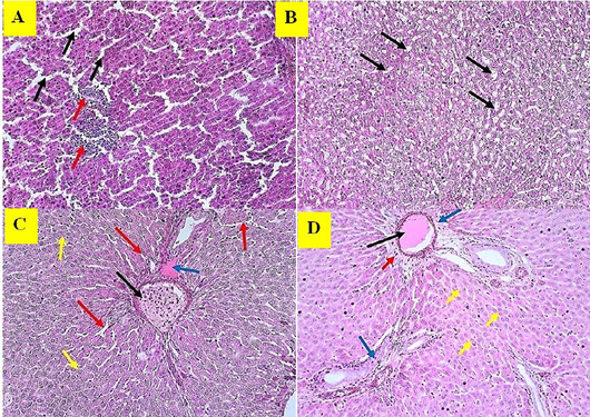
Figure 13:
Histopathological changes of liver in mice received estrogen. (A): Sinusoidal dilation (Black arrow) and infiltration of mononuclear cells (Red arrow); (B): Droplets of cytoplasm (Black arrow); (C): Congestion of veins (Black arrow), Amyloidosis (Blue arrow), Sinusoidal space (Red arrow), Degeneration of parenchyma (Yellow arrow); (D): Thrombus (Black arrow), Deposition of fibrin (Blue arrow), Perivascular cuff (Red arrow), Degeneration of parenchyma (Yellow arrow). The sections were stained with Hematoxylin and Eosin stain, examined at 10X, and the images are represented experimental mice.