
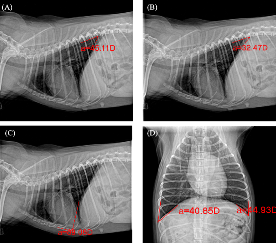
Fig. 1.
Imaging of PLFA. A, VAcr on the right lateral view. B, VAca on the right lateral view. C, SA on the right lateral view. D, CAl and CAr on the ventral-dorsal view.
Imaging of PLFA. A, VAcr on the right lateral view. B, VAca on the right lateral view. C, SA on the right lateral view. D, CAl and CAr on the ventral-dorsal view.