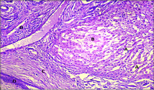
Figure 7:
Histopathological section of liver showing; A: dilated sinusoids; B: coagulative necrosis of hepatocytes; C: fibrosis of portal area; D: migratory tract with inflammatory cell infiltration. (H & E stain. X20).
Histopathological section of liver showing; A: dilated sinusoids; B: coagulative necrosis of hepatocytes; C: fibrosis of portal area; D: migratory tract with inflammatory cell infiltration. (H & E stain. X20).