
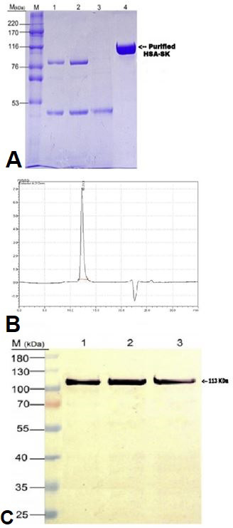
(A) SDS-PAGE analysis of purified HSA-SK as a result of one step chromatographic purification by capture select human serum albumin affinity marix. Here Lane M represents protein markers of high molecular weight, Lane 1 contain proteins from broth supernatent, Lane 2 contain protein from diafiltered supernatent, Lane 3 represents protein from flow through and finally, Lane 4 represents the final eluted fusion protein HSA-SK. (B) Chromatogram is showing the analysis of purified HSA-SK fusion protein by RP- HPLC. 210 nm detector was used for the absorbance of protein and chromatogram showed a single sharp peak at retention time 10.7 min. (C) Western blot analysis of purified fusion protein HSA-SK where Lane M contain marker protein, Lane 1 represents a band of purified HSA-SK, Lane 3 has a band of non-reduced HSA-SK and Lane 3 shows a band of HSA-SK standard.