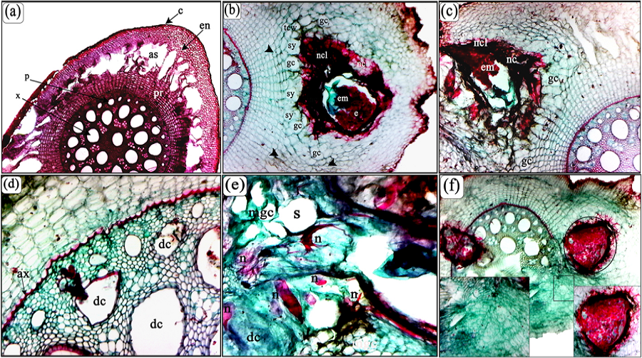
(a). Transverse section of healthy banana cv. Hindi roots; Central cylindrical or stele [Phloem (p), xylem (x)], Prosenchyma (pr), Endodersmis (en), Mesodermis (parenchyma) (m), Cortex (Epidermis and exodermis) (c), Air space (as). (b) Transverse section through the gall and cortex cells of the infested banana root with Meloidogyne incognita showed the feeding site of adult female with egg masses (em), note the multinucleate cells, giant cells and lateral expansion in necrotic cell (nc) within layers (lnc) in the inner cortical tissue is beginning to spread to the stele with compressed of cells size in various layers of the of the root (head arrow) with thickness in cell walls (tcw). (c) showing lateral expansion of giant cells (gc) necrotic cell (nc) within layers (lnc) in the inner cortical tissue is beginning to spread to the stele through a passage cell in the endodermis (e). (d) The deformed cells in the parenchyma and abnormal xylem (ax). (e) Fourth stage of M. incognita (n) near of the cambium (ca). It notes a large laceration in neighboring cells of feeding site; multinucleate giant cells (mgc), note dark tissue elements and thickness of the cell wall in neighbor cells of multinucleate cells and dense cytoplasm. (f) Adult female and eggs of M. incognita near of the vascular cylinder, notes a large laceration in neighboring cells of feeding site. (Scale bars: a-f = 200 μm).