
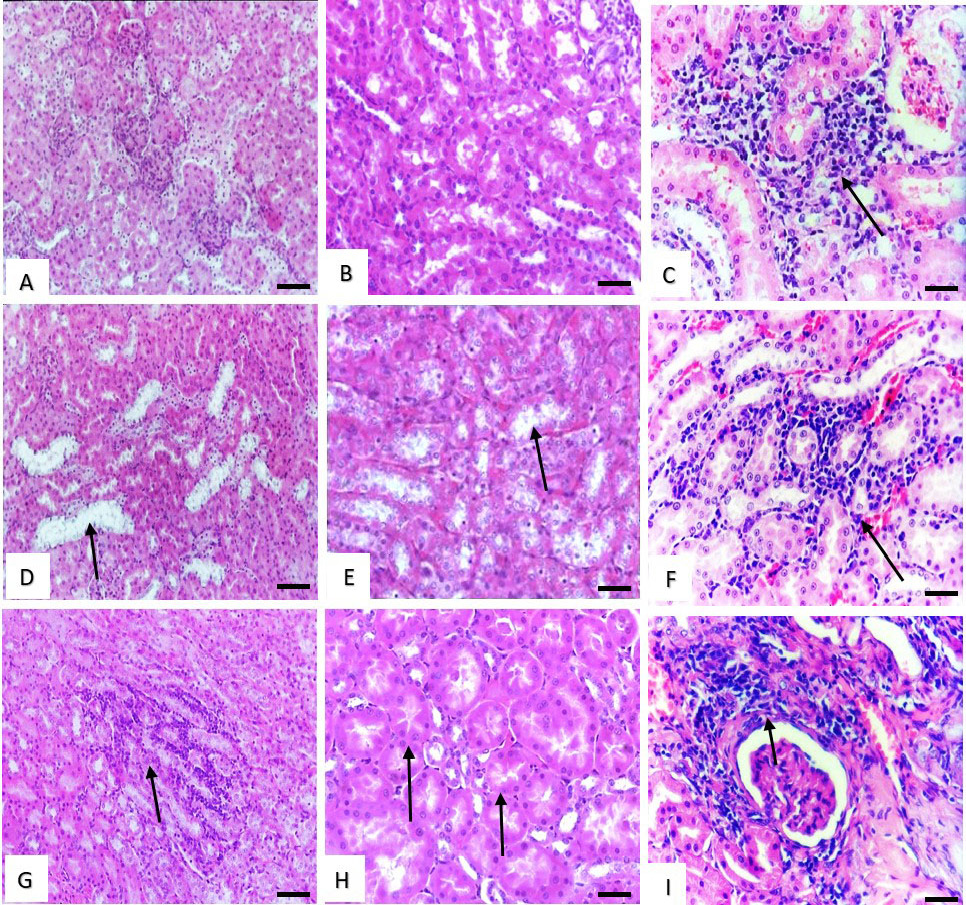
Fig. 3.
Histological structure of rat kidney. (A and B) Effect of low dose of malathion and metalaxyl showing normal structure. (C) Effect of low dose of cymoxanil showing, slight interstitial mononuclear cells infiltration (arrow). (D and E) Effect of medium dose of malathion and metalaxyl showing slight vacuolar degenerative changes in the renal tubules lining epithelium (arrows). (F) Effect of medium dose of cymoxanil showing slight interstitial nephritis (arrow). (G) Effect of high dose of malathion showing marked interstitial nephritis (arrow). (H) Effect of high dose of metalaxyl showing cloudy swelling in the epithelial lining of the renal tubules (arrows). Stain: H and E. Magnification bar: 50 µm.