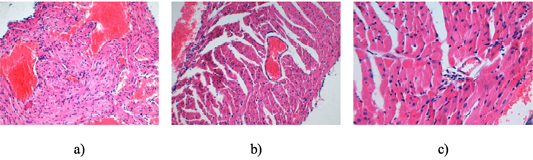
Figure 4:
a) Cardiac myocytes of the control group animal H and E staining. 10Xoc.lens, 10Xobj.lens; b) Cardiac myocytes of the animal from the experimental group 1. H and E staining. 10Xoc.lens, 4Xobj.lens; c) Heart muscle tissue of animals given pantocrine. H and E staining. 10Xoc.lens, 20Xobj.lens.