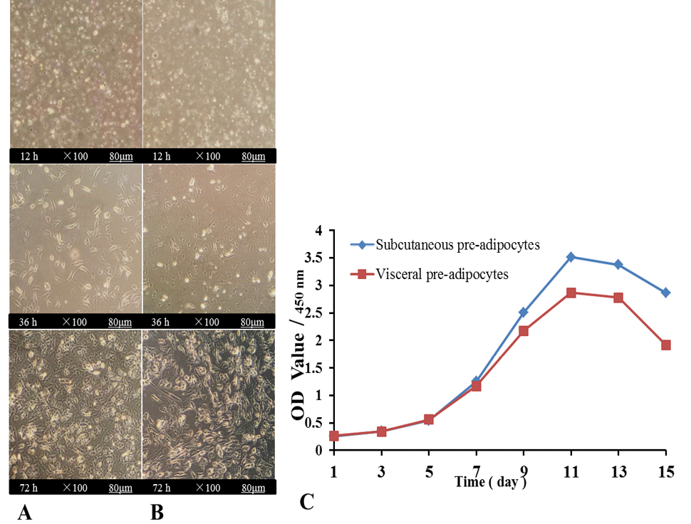
Fig. 1.
Morphology of pre-adipocytes of subcutaneous and visceral extracted in vitro. The morphology of cells at 100 ×, A and B scalebar was 80 μm. Subcutaneous pre-adipocytes (A) and visceral pre-adipocytes (B) were photographed under the bright-field microscope at the indicated time (12 h, 36 h, 72 h). Mouse subcutaneous and visceral primary pre-adipocytes adherent cells were observed 12 h after inoculation, which were quasi-round in shape and varied in size. After 36 h of inoculation, the number of adherent cells increased greatly, and the shape was fusiform, polygonal or irregular, with a strong three-dimensional feeling. About 72 h after inoculation, the primary cells can grow to monolayer fusion, and then the primary cells become long fusiform and oval in further culture. (C) The growth curve of mouse primary subcutaneous and visceral pre-adipocytes at the indicated days (D0, D1, D3, D5, D7, D9, D11, D13, and D15). Values are presented as the mean ± SD.