
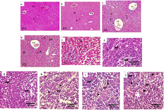
Figure 4
Hematoxylin–eosin-stained histological sections of the maternal liver (a-e) and spleen (f-j). For the liver, (a,b) normal hepatocyte devoid intracellular gaps; (c,d, e) infiltration of leukocyte and deposition of malarial pigment (H and E stained, 100x). (a) non pregnant, (b) pregnant, (c,d,e) pregnant-infected. For the spleen (f, g) normal texture of splenocytes; (h, i, j) expanded of red pulp along with the deposition of malarial pigments (H and E stained, 400x). (f) non-pregnant, (g) pregnant, (h, i, j) pregnant-infected. VS: central vein; VP: portal vein; S: sinusoid; N: necrosis; I: leukocyte infiltration; K: Kupffer cells; P: malaria pigmentation; WP: White pulp; RP: red pulp; Arrowheads: Hemozoin.