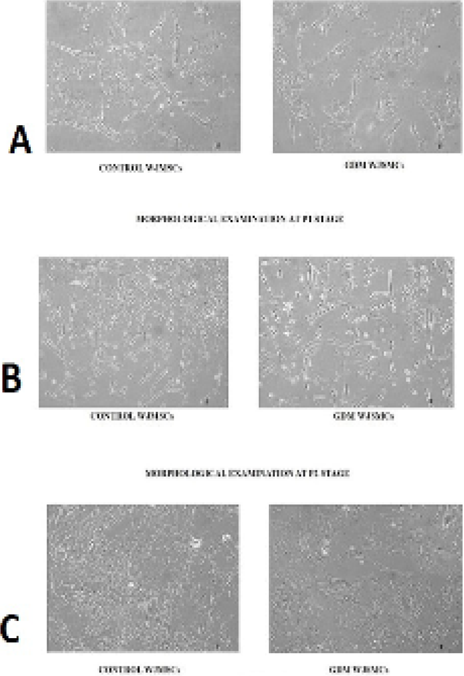
Fig. 2.
Morphological examination of control and GDM WJMSCs at P0, P1, P2 (passage 0, 1, 2) stage and magnification power, under inverted microscope (Floid Cell Imaging System). WJMSCs of both groups were analyzed under inverted microscope at different passaging stages (P0, P1, P2) at 10x magnification power. At P0 stage WJMSCs showed large spindle shaped morphology compared to control WJMSCs (A). At P1 stage GDM WJMSCs appeared as flattened as well as spindle shaped as compared to control (B). While at P2 there was no difference seen in morphology of WJMSCs in both groups showed normal spindle shaped morphology (C).