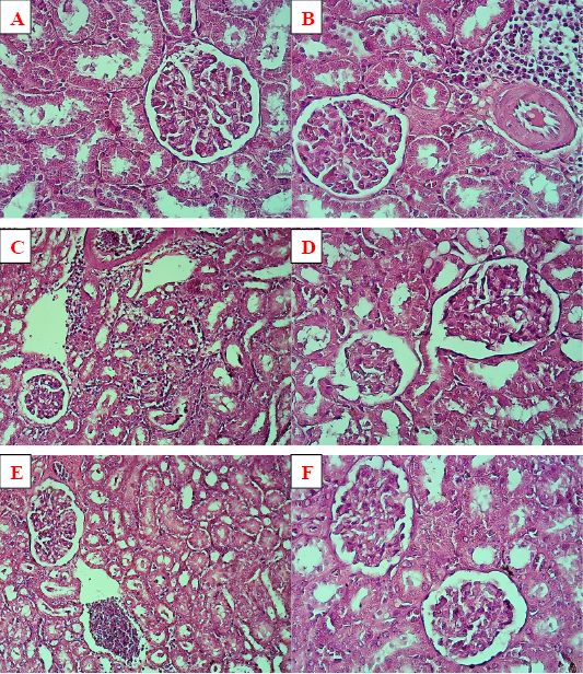
Figure 2
Histopathology of renal tissue. (A) negative control with normal glomeruli and renal tubules, X40. (B) positive control with aggregate of inflammatory cells among renal tubules and vascular congestion, X40. (C) and (D) rousovastatin group with massive infiltration of inflammatory cells in renal tissue along with some degenerative changes of renal tubules and atrophy of glomeruli, X20, X40 respectively. (E) combination group showed congestion and vascular aggregate of inflammatory cells with a lesser degree, X20. (F) pumpkin group showed a normal renal tissue, X40.
