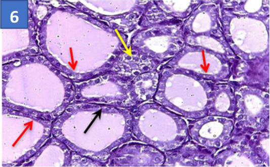
Figure 6:
Section of thyroid gland treated by SAL+ALA: Showing more improvement thyroid follicle wall lined with typical cuboid epithelial cells (red darts), with clear parafollicular cells (yellow darts), thin septa of collagen fibers between thyrocytes and follicles (black dart) (H & E) stain(40X).