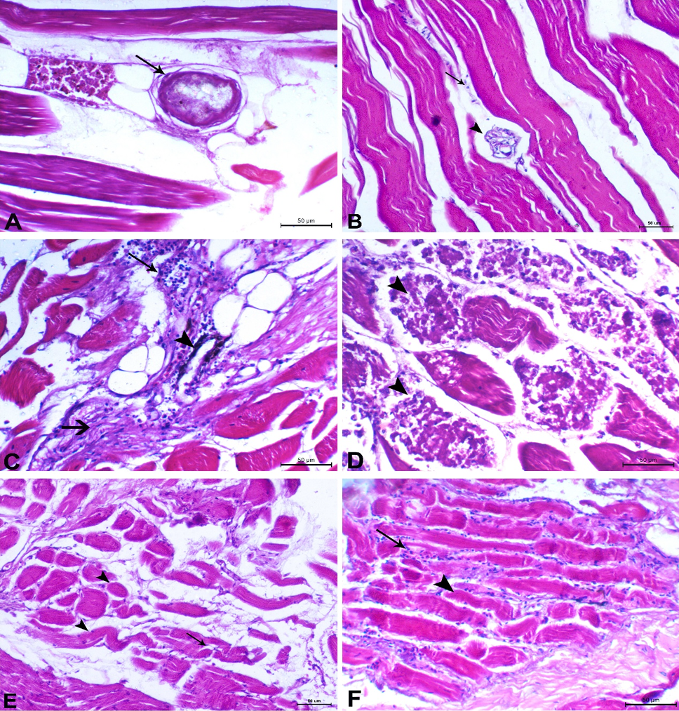
Histopathological findings of EMC infection in muscles of tilapia and mullet fish samples. A, Mullet muscle showing EMC embedded in the intermuscular fat (arrow). B, Nile tilapia muscle showing EMC (arrowhead) between the muscle bundles with few mononuclear cells infiltration (arrow). C, Mullet muscle showing marked degenerative changes of the muscles associated with mononuclear cells infiltration (arrow) and melanophores(arrowhead) with foal areas of muscular hyalinization (thick arrow). D, Mullet muscle showing marked necrotic changes of the muscle bundles with lysis of myofibers (arrowheads). E, Mullet muscle showing atrophy of muscle bundles (arrow heads) associated with destruction and loss of muscle bundles irregularities (arrow). F, Tilapia muscle showing atrophy of muscle bundles (arrowhead) accompanied with mononuclear cells infiltration (arrow). Stain H and E, x200, scale bar 50μm.
